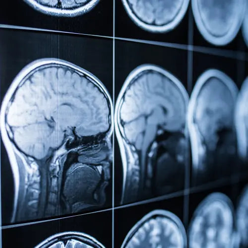
The brain does not hurt! It is not sensory innervated. The pain comes from its envelope called the meninges. Here is a short description of this structure that makes migraine sufferers suffer so much.
Brains
has. Anatomy of the dura mater
The meninges are membranes that envelop the central nervous system , the brain and the spinal cord, the intracranial portion of the cranial nerves and the roots of the spinal nerves. Their main function is to protect the nervous system from shocks . They also supply nutrients to the nervous system and play a role in brain immunity . We distinguish, from the surface towards the depth, the dura mater, the arachnoid and the pia mater .Classically, the anatomical conception of the meninges admits that these are three in number, but recently, a more modern conception tends to speak of meninges with 2 layers, a soft meninge and a hard meninge .
The soft meninge is the joining of the pia mater and the arachnoid which we gather under the term of leptomeninge . It is a loose tissue, intermediate between the dura mater and the neuraxis. Cerebrospinal fluid is found between these layers and in the subarachnoid space.
The dura mater is the hard meninge whose main roles are support and protection . It is made up of thick layers of fibrous, elastic tissue that are attached to the bones above. It is divided into an epidural layer which adheres to the bones of the skull and a meningeal layer in direct contact with the soft meninges. The two layers are laid one above the other, with no space between them.
The inner surface of the dura mater is lined with the parietal leaflet of the arachnoid , which adheres strongly to it. From this surface is detached a certain number of prolongations or partitions which are interposed between the various segments of the encephalic mass . This makes it possible to isolate the compartments from one another and thus to ensure that their situation is maintained, whatever the position of the head. There are four partitions which are the tent of the cerebellum, the falx of the brain, the falx of the cerebellum and the tent of the pituitary gland (Marieb 2014).
 Meninges as central part of brain structure or under skin layers outline diagram Stock Vector by ©VectorMine 517281978. Depositphotos
b. Vascularization of the meninges
The meningeal arteries that supply the dura mater and partly the adjacent bones come from the internal and external carotid systems and form an anastomotic network on the surface of the dura mater. They consist of the anterior meningeal artery , which originates from the ophthalmic or ethmoidal artery ; the middle meningeal artery which originates from the internal maxillary artery ; the meningeal branches of the intracavernous internal carotid artery and the posterior meningeal artery which originates at the level of the occipital or vertebral artery (Marieb and Hoehn 2014).
vs. Innervation of the meninges
The meninges and their vascular network are innervated by the sympathetic , parasympathetic and sensory fibers. The supratentorial level is innervated by branches of the trigeminal nerve (V) and the infratentorial level by the superior cervical nerves and the vagus nerve (X) . For a long time, sensory innervation was attributed to the dura mater, but it has now been demonstrated that the other layers of the meninges, and in particular the pia mater and its small vessels, can also be mediators of pain . (Dallel et al. 2003; Fontaine et al. 2018). This nociceptive innervation more specifically involves the trigeminal afferents which, through the superior salivary nucleus, are connected to the neurovegetative efferents (May et al. 1999).
We recommend the article: Mechanism of migraine

Dallel, Radhouane, Luis Villanueva, Alain Woda, and Daniel Voisin. 2003. “Neurobiology of trigeminal pain”. Medicine/Science 19 (5): 567-74. https://doi.org/10.1051/medsci/2003195567.
Fontaine, Denys, Fabien Almairac, Serena Santucci, Charlotte Fernandez, Radhouane Dallel, Johan Pallud, and Michel Lanteri-Minet. 2018. “Dural and Pial Pain-Sensitive Structures in Humans: New Inputs from Awake Craniotomies.” Brain 141 (4): 1040‑48. https://doi.org/10.1093/brain/awy005.
Marieb, Elaine, and Katja Hoehn. 2014. Human Anatomy and Physiology . Pearson Education France. May, Arne, and Peter J. Goadsby. 1999. “The Trigeminovascular System in Humans: Pathophysiologic Implications for Primary Headache Syndromes of the Neural Influences on the Cerebral Circulation.” Journal of Cerebral Blood Flow & Metabolism 19 (2): 115-27. https://doi.org/10.1097/00004647-199902000-00001.


